Figure 2.

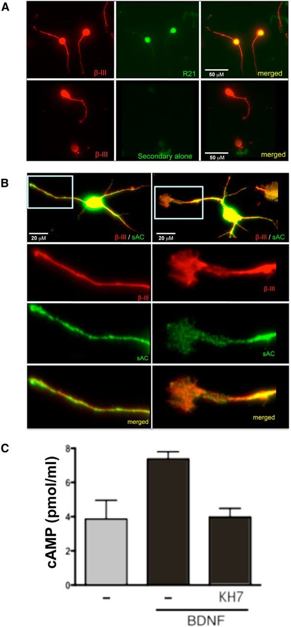
Expression of sAC in rat postnatal primary neurons. A, Dissociated CGNs were labeled with anti-β-III-tubulin (red) and either anti-sAC (R21; green, top) or secondary antibody alone (green, bottom). Scale bars: 50 μm. B, Dissociated cortical neurons were labeled with anti-β-III-tubulin (red) and anti-sAC (R21; green). Shown below are higher magnifications of the boxed neurite (left) and growth cone (right). Scale bars: 20 μm. C, Accumulated cAMP was measured in CGNs after 2 min of stimulation with BDNF (200 ng/ml) in the presence of IBMX (1 mm), ±KH7 (50 μm). Shown are averages of triplicate determinations of a representative experiment (repeated three times).
