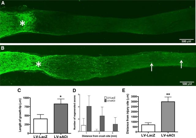
Figure 6.
Overexpression of sAC promotes axonal regeneration in vivo. The right optic nerves of adult Fisher rats (200–250 g) were exposed and crushed 2 mm behind the eye. LV-sACt or LV-LacZ were injected into the vitreous chamber of the right eye. Animals were killed after a 2 week postsurgical survival period, then optic nerves were sectioned and immunostained for GAP-43. Each injection was done without injuring the lens, which if injured, could cause inflammation and inadvertently result in promoting optic nerve regeneration (Leon et al., 2000). Representative images from an LV-LacZ (A) and a LV-sACt (B) infected animal. Arrows illustrate regenerating axons. Asterisk (*) indicates the injury site. C, The distance of the growth tip of robust growth, and (D) the average number of regenerated axons at 1, 2, and 3 mm beyond the injury site were measured for each fiber. E, The average distance of regeneration beyond the injury site was measured for the three longest axons for each animal, 5–7 animals per group; *p < 0.05, **p < 0.01. Scale bars, 500 μm.

