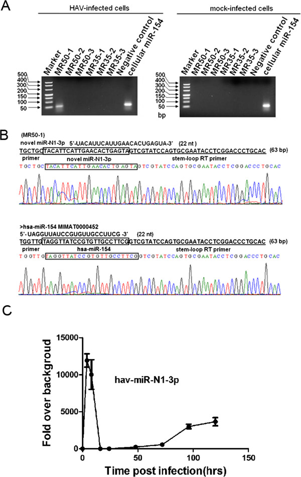
Figure 3.

Cloning and expression validation of the predicted miRNAs in HAV-infected and mock-infected cells. (A) A HAV antigenome-encoded miRNA was detected in the HAV-infected cells. 4% agarose gel electrophoresis analysis showed stem-loop RT-PCR products with approximately 63 bp specific bands. Cellular miR-154 was used as a positive control when performing agarose gel electrophoresis. (B) Sequencing results for the novel HAV miRNA and primer sequences are underlined in the sequencing maps. (C) Time course analysis of hav-miR-N1-3p in KMB17 cells. KMB17 cells were infected with HAV at a MOI of 0.5 TCID50/cell. Total RNA was extracted at 0, 4, 8, 12, 24, 48, 72, 96 and 120 hpi. and measured by qRT-PCR to quantify the expression of hav-miR-N1-3p during infection. All values are presented as changes in expression relative to uninfected cells after normalization to values of snU6 RNA.
