
Figure 3.
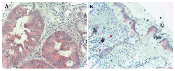
EGFR immunohistochemistry in CRC. A: Carcinomatous glands of the colon showing diffuse cytoplasmatic, moderately intensive EGFR staining (Hematoxylin co-staining × 200); B: Normal colonic epithelia showing mildly, moderately intensive basolateral intracytoplasmatic EGFR staining. The lower 2/3 of the crypts do not show EGFR positivity (Hematoxylin co-staining × 100).