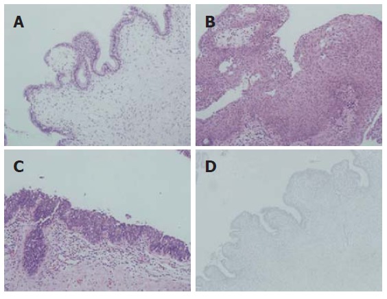
Figure 2.

Microscopically, the cyst wall showing a variety of lining epithelia including tall columnar or cuboidal (A), squamous (B), and transitional (C) epithelium (HE, x 200), the underlying stroma showing a mild infiltrate of chronic inflammatory cells, immunohistochemical staining showing negative prostate specific antigen in the lining of epithelial cells (D) (PAP, x 200).
