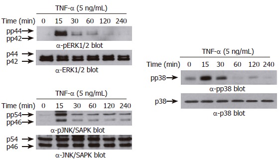
Figure 5.

TNF-α induces phosphorylation of MAPKs in AGS cells. AGS cells (5 x 10⁵ cells/well) were incubated for various times (0-240 min) with TNF-α (5 ng/mL). Protein extracts were prepared at the indicated time points, and then the levels of phosphorylated or total MAPKs (ERK-1/2 (top), p38 kinase (middle), and JNK/SAPK (bottom)) were determined by Western blotting using specific antibodies. The arrows indicate the position of specific immunoreactive bands corresponding to distinct MAPKs.
