
Figure 3.
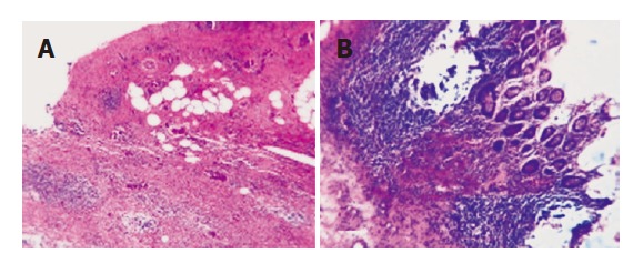
Xanthogranulomatous cholecystitis (A) and xanthogranulomatous cholecystitis involving the wall of the transverse colon (B). There is destruction of the submucosa and muscular coat of the transverse colon by extensive macrophage infiltrates, the mucosa is intact and exhibits no cellular atypia (HE, 200x).