Figure 3.

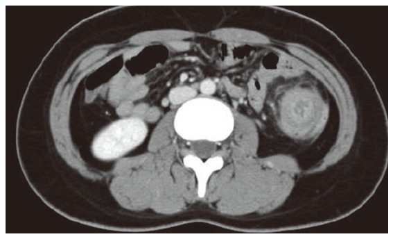
Computed tomography shows the cystic lesion in the descending colon, occupying the whole lumen, and also shows multicentric target sign and sausage-shaped inhomogeneous soft-tissue mass.

Computed tomography shows the cystic lesion in the descending colon, occupying the whole lumen, and also shows multicentric target sign and sausage-shaped inhomogeneous soft-tissue mass.