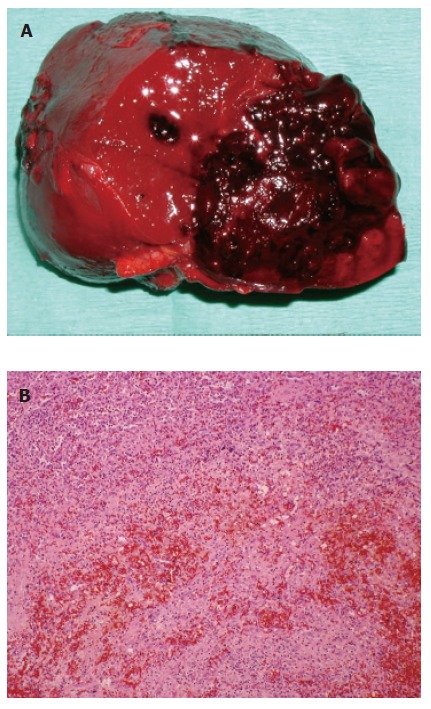
Figure 2.

A: Macroscopic findings for the ruptured splenic hamartoma. The hamartoma is well demarcated from the surrounding parenchyma and significant hemorrhage is shown; B: Microscopy showed a red pulp with no lymphatic structures, or trabeculae of the spleen. (hematoxylin-eosin, original magnification, ×200).
