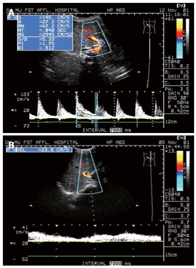
Figure 1.

A: Duplex Doppler Sonogram of case with IHS showed the PHA was easy to see as well as PSV and RI of the PHA increased significantly; B: Duplex Doppler Sonogram of the normal subject showed normal waveform of PV, and PHA cannot display clearly.
