
Figure 1.
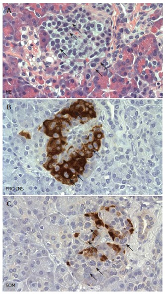
Histopathological features of diffuse nesidioblastosis. A: HE stained sections demonstrating a prominent lobulation of an islet. Some of the endocrine cells showing hyperchromatic and enlarged nuclei are labelled with arrows. B, C: Adjacent section analysis demonstrating cytoplasmic positivity for proinsulin (PRO-INS) in those endocrine cells with hyperchromatic nuclei (arrows in B). In contrast, these cells are negative for somatostatin (SOM) (arrows in C).