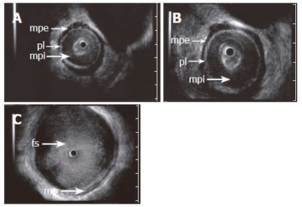
Figure 2.

ES images (A and B) obtained with a 15 MHz miniprobe in a patient with achalasia showing the longitudinal, outer layer of the proper muscle (mpe) separated from a thickened circular, inner layer (mpi) by an echogenic layer corresponding to the localization of the plexus myentericus (pl). C: shows a dilated esophageal lumen with food content (fs). A normal aspect of the proper muscle is seen.
