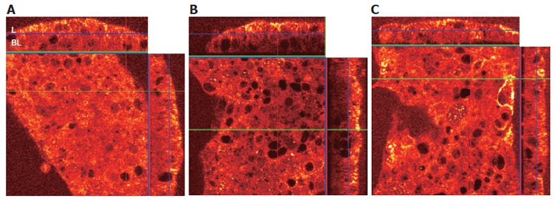
Figure 4.

A: Viewing the X-Z panels, the FADD (stained red, with higher densities staining yellow) in untreated (control) cells distributed equally throughout the cytoplasm. “L” represents the apical (lumenal) surface and “BL” represents the basolateral surface of the N87 monolayer; B: Labeled FADD in cells treated with the Fas trimerizing antibody is recruited to the apical surface, leaving much of the cytoplasm with reduced FADD staining; C: When the anti-MHC class II IgM RFD1 is applied to the cell monolayer 30 min prior to treatment with the Fas agonist IPO4 IgM, the recruitment of FADD to the cell surface is markedly reduced.
