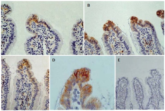
Figure 2.

Immunohistochemical expression of claudin-4 in the mucosa of terminal ileum: In control and sham operated rats claudin-4 is expressed on the basolateral sides of a few villous surface epithelial cells (A: original magnification x 400). In obstructive jaundice, immunoreactivity for claudin-4 is stronger and this molecule is expressed by most epithelial cells lining throughout the upper third of the villi and not only in the tip (B: original magnification x 400). At higher magnification we can see the enhancement of the lateral distribution of claudin-4 in obstructive jaundice (D: original magnification x 1000). In bombesin or neurotensin-treated rats claudin-4 expression is restored to the control level (C: original magnification x400). Substituting anti-claudin-4 antibody by mouse serum no staining is observed (E: original magnification x 200).
