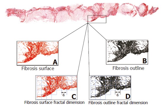
Figure 1.

Fractal morphometry of liver fibrosis in two-dimensional biopsy sections. The software automatically selects the fibrosis surface (A) on the basis of RGB colour segmentation, and its outline perimeter (B) whose calculated length was subsequently used to estimate the wrinkledness index. The fractal surface (C) and outline (D) dimensions of fibrosis were estimated using the box-counting algorithm. Covering an irregular surface requires the intersection of contiguous and non-overlapping two-dimensional boxes with a side length of ε: We counted the number of boxes that contain at least one point of the object N(ε) for different box sizes ε and then determined the fractal dimension (DB) by using Equation (1).
