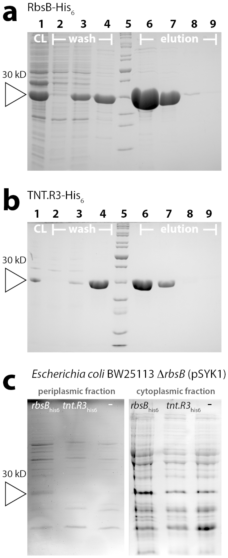
Figure 2. Purification and expression in E. coli of RbsB-His6 and TNT-His6.

(a) Coomassie-stained SDS-PAGE gel of purified fractions of RbsB-His6 from E. coli BL21 (pAR1, strain 3725). Lanes: 1, cleared lysate; 2, flowthrough; 3, Washing step I; 4, Washing step II; 5, Peqgold protein marker 2; 6, Elution I; 7, Elution II; 8, Elution III; 9, Elution IV. (b) As A, but for E. coli BL21 (pAR2, strain 3325). Lanes: as for panel (A). (c) InVision His6-tag stained SDS-PAGE gel of cell extracts from E. coli BW25113 ΔrbsB (pSYK1) expressing rbsB-his6 (from plasmid pAR3, strain 4175), tnt.R3-his6 (from plasmid pAR4, strain 4176), or with empty pSTV plasmid (strain 4497). Left panel, periplasmic protein fraction. Right panel, whole soluble protein fraction. Open triangle indicates the expected position (30 kDa) of RbsB-His6 and TNT.R3-His6.
