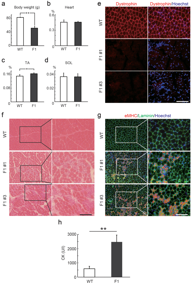
Figure 3. Inheritance of mutations in the rat Dmd gene generated by CRISPR/Cas9.
Comparison of body weight (a), weights of the heart (b), tibialis anterior (TA) muscle (c), and solaris (SOL) muscle (d) relative to body weight of 4-week-old wild type (WT) and F1 male rats. *P < 0.05, **P < 0.01. (e) Cross sections of TA muscles in 4-week-old WT and F1 male rats stained with anti-Dystrophin (red) antibody. Scale bar = 100 μm. (f) Hematoxylin and eosin (H&E) staining of TA muscles in 4-week-old WT and F1 male rats. Scale bar = 100 μm. (g) Cross sections of TA muscles in 4-week-old WT and F1 male rats stained with anti-laminin (green) and anti-embryonic myosin heavy chain (eMHC, red) antibody. Scale bar = 100 μm. (h) Creatine kinase (CK) activity of Dmd-mutated F1 (n = 5) and age-matched WT (n = 3) rats. **P < 0.01.

