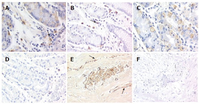
Figure 1.

Expression of Kit in normal gastric tissue and gastroparesis (x 200). In both normal (A) and gastroparetic stomach (B), Kit was detected on mast cells (arrows) used as an internal control; In normal gastric samples, positivity is shown on parietal cells (C) and at the intrinsic innervation level (E, arrows), whereas in gastroparesis no staining is present at either levels (D, F).
