Figure 1.

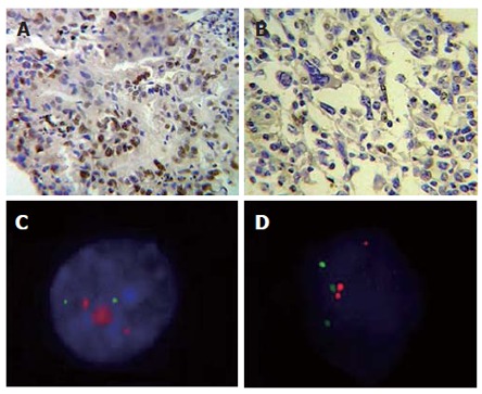
Cells submitted to immunohistochemistry and FISH techniques. A: Infiltrating gastric adenocarcinoma of intestinal type shows intense nuclear marcation for C-MYC, × 400; B: Gastric adenocarcinoma of diffuse type nuclear marcation and cytoplasmatic light marcation for C-MYC, × 400; C: Interphase nuclei presenting C-MYC high amplification (red) and chromosome 8 (green); D: Interphase nuclei presenting chromosome 8/C-MYC rearrangement.
