
Figure 3.
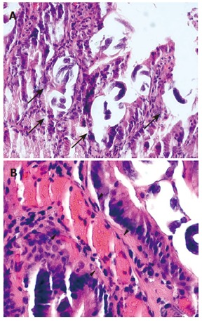
A: Intraluminal and intramucosal S. stercoralis larvae in the duodenum during the exudative enteropathy hyper-infection phase (arrows). Scale: 200 μm. B: No intramural or peri-vascular inflammatory cell clusters are recognised during this early period of infection. Scale: 50 μm.