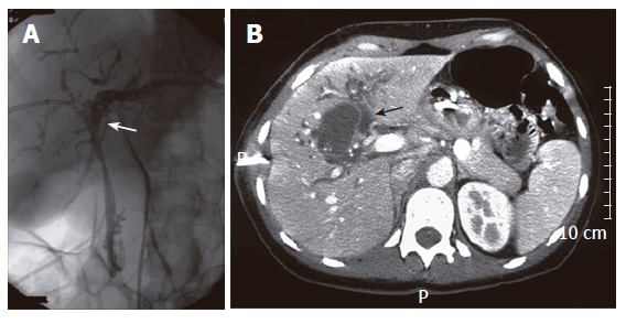
Figure 4.

Percutaneous transhepatic drainage (PTD) showing complete obstruction at level of the proximal bile duct (A), abdominal CT showing a cystic lesion with irregularly thickened wall in conjunction with dilatated intrahepatic bile ducts (arrow) (B) in case 5.
