
Figure 3.
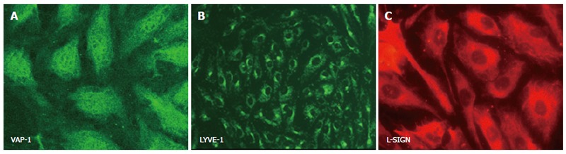
Cultured human hepatic sinusoidal endothelial cells stain positively with antibody directed against ‘non-classical endothelial’ phenotypic markers. Images represent immunofluorescent staining of cultured cells using specific primary antibodies in an indirect fluorescent protocol. Expression of VAP-1 (A) and LYVE-1 (B) are visualised with a FITC-conjugated secondary antibody (green), whilst positive staining for L-SIGN (C) is visualised using a Texas Red-labelled secondary antibody.