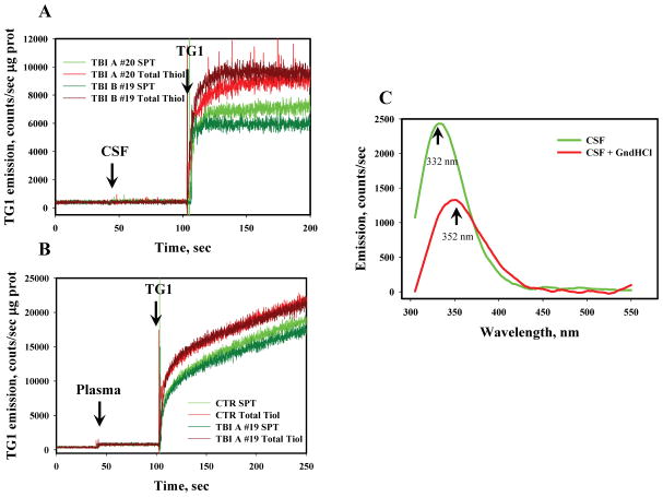
Fig. 1. Analysis of total- and protein-thiols in CSF and plasma of control and TBI patients.
Panel A: kinetics of TG-1 (final concentration 5 μM) emission with TBI CSF samples (#19 and 20) diluted (1:100) in 20 mM phosphate buffer (pH=7.4, 37°C) before and after SEC with BS6. Panel B: kinetics of TG-1 emission after plasma sample (TBI #19 and control) dilution (1:100) in 20 mM phosphate buffer (pH=7.4, 37°C) before and after SEC with BS6. Panel C: Effect of GnHCl (~5.4 M) on CSF protein denaturation through analysis of protein tryptophanyl fluorescence (Ex. 295 nm). Data are representative of 3 independent experiments.

