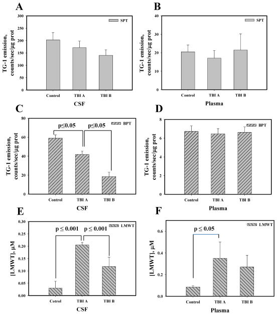
Fig. 2. Redox status of thiols in CSF and plasma of control and TBI patients.
Panels A and B: surface protein thiol (SPT) in CSF and plasma, respectively. Panels C and D: buried protein thiols (BPT) in CSF and plasma, respectively. Panels E and F: low molecular weight thiols (LMWT, mainly GSH) in CSF and plasma, respectively. CSF and plasma matching samples were collected at the time of EVD placement (TBI A) and 24 hours later (TBI B). TG-1 emission was normalized for protein content. For LMWT TG-1 emission was recalculated into μM concentration using linear calibration curve with GSH. Data presented as mean±SD for 3 independent experiments.

