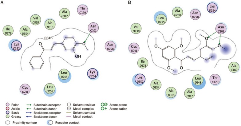
Figure 10.
Ligand-protein interaction diagrams for the binding site of tubulin with two hit compounds. The hits are: (A) compound 1 (AE-562/40322474) (B) compound 3 (AQ-358/41842921). Residues are annotated with their 3-letter amino acid code. There are five chains in the system and its positions are prefixed with letters of the alphabet. Hydrogen-bonding interactions between the receptor and the ligand are drawn with an arrowhead to denote the direction of the hydrogen bond. When the hydrogen bond is formed with the residue sidechain, the arrow is drawn in green.

