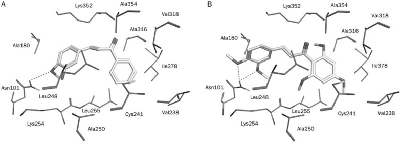
Figure 11.
Molecular docking results. Docked orientations of (A) compound 1 (AE-562/40322474) (B) compound 3 (AQ-358/41842921). The active site residues are shown in stick form. Hydrogen-bond network with protein residues is represented in red dotted lines. All the compounds are color-coded by yellow.

