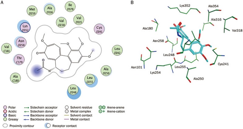
Figure 6.
Interaction analysis. (A) 2D interaction diagram for the binding site of tubulin with the colchicine. Residues are annotated with their 3-letter amino acid code. There are five chains in the system and its positions are prefixed with letters of the alphabet. (B) 3D interaction diagram for the binding site of tubulin with the colchicine. The active site residues are shown in stick form. The colchicine is color-coded by cyan.

