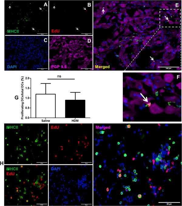
Figure 7.

Quantitative analysis of proliferating DCs in JNC. A: MHC II staining of DCs. B: EdU detection of proliferating DCs (arrows). C: Nucleus staining with DAPI. D: Neurons positive for PGP 9.5. E: Merged representation of the images shown in A-D. F: An enlargement of EdU positive cell. G: Quantification of proliferating DCs in JNC of HDM treated and control mice. Ganglion sections were stained for EdU, MHC II, PGP 9.5 and nucleus. The proliferating DCs, which are positive with MHC II and EdU labelled nucleus, and total number of DCs were evaluated. The results were expressed as percentages of proliferating DCs/total DCs. The proliferation rate of DCs in HDM treated mice 0.89 ± 0.38% (n = 4) and control mice 1.19 ± 0.54% (n = 5), p = 0.68. H: The positive control of EdU in the lung. The figures showed amount of proliferating MHC II-IR cells and other cell types in the lung. Scale bars: 100 μm in A, B, C, D, 50 μm in H.
