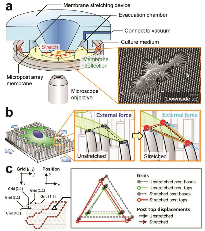
Figure 1.
Live-cell subcellular measurement of cell stiffness using a microengineered stretchable micropost array membrane (mPAM) and a cell stretching device (CSD). (a) Schematic of the CSD and its implementation for stretching the mPAM and thus single cells adhered on the tops of the PDMS microposts. Only one-half of the CSD is shown for visualization of its internal structure. A vacuum was applied to the CSD evacuation chamber to draw the periphery of the mPAM, causing the central region of the mPAM holding the PDMS micropost array and pre-seeded with adherent cells to stretch equibiaxially. Inset: Scanning electron microscopy image of a single cell seeded on the PDMS micropost array. Scale bar: 20 μm. (b) Cartoon of an adherent cell on the mPAM under a static equibiaxial stretch. Left inset shows a triangular subcellular region formed between three adjacent microposts deflected by cellular traction forces. Right inset shows the same triangular region under a static equibiaxial stretch with an increased cell area and heightened traction forces. Mechanical forces exerted on the cell by the PDMS microposts were labelled by blue and light blue arrows before and after the mPAM stretch, respectively. (c) Grid arrangement for theoretical computation of subcellular cell stiffness. Hexagonally arranged microposts formed the nodes in a regular triangular grid surface. Inset: Relative displacements of the micropost tops before and after static equibiaxial mPAM stretches used to calculate stretching forces and deformations of grid elements.

