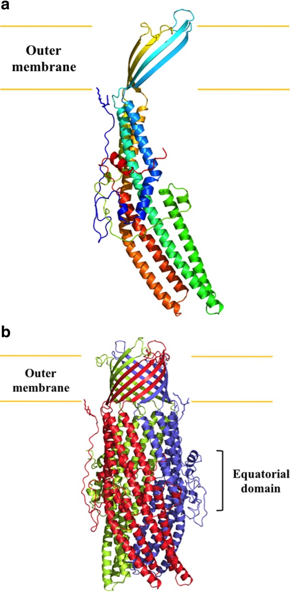
Figure 2.

Structure of the C. jejuni CmeC channel protein. (a) Ribbon diagram of a protomer of CmeC viewed in the membrane plane. The molecule is colored using a rainbow gradient from the N-terminus (blue) to the C-terminus (red). (b) Ribbon diagram of the CmeC trimer viewed in the membrane plane. Each subunit of CmeC is labeled with a different color. The CmeC protomer is acylated (in sticks) through the first N-terminal cysteine residue to anchor onto the outer membrane.
