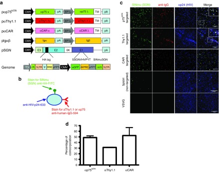
Figure 1.

Characterization of targeted lentiviral vectors. (a) Schematic drawing of plasmids used for production of targeted vectors. Targeted vectors were generated by six-plasmid cotransfection using the (i) pαThy1.1/pαp75NTR/pαCAR plasmids, (ii) Igαβ plasmids required for surface expression of antibody molecules (iii) SINmu(SGN) envelope plasmid, the (iv) pRRLsincppt_CMV_eGFP-WPRE genome plasmid and the pMD2-LgpRRE and pRSV-Rev packaging plasmids. (b) Schematic representation of the virus staining method for visualizing individual particles. (c) αp75NTR, αΤhy1.1, αCAR, 5pl, and VSVG particles were overlaid upon coverslips and stained for the presence of SGN fusogen (anti-HA-FITC), αp75NTR/αThy1.1/αCAR (Alexa 594 anti-human IgG) and HIV p24 (anti-p24). The insert shows zoomed in view of the indicated region. Colocalizing pixels are indicated by a white signal. Bars = 10 μm. (d) Quantification of triple positive particles. For α75NTR 49.8% of particles codisplay antibody and fusogen molecule, while for αThy1.1 and for αCAR 31 and 53% of total particles are respectively triple positive. CAR, coxsackievirus and adenovirus receptor; CMV, cytomegalovirus; EGFP, enhanced GFP.
