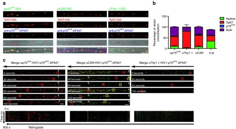
Figure 5.

Retrograde axonal transport of targeted vectors. (a) Primary motor neurons plated in microfluidic chambers were incubated with either DiO labeled αp75NTR or αThy1.1 or αCAR with TeNT-AF555 and anti-p75NTR-AF647 for 2 hours at 37 °C before fixing and imaging by confocal microscopy. Shown are representative maximum projections of z-stack images where white circles show examples of colocalization of vectors with both markers and yellow circles show examples of colocalization with just TeNT-AF555 and green circles show non-associated vectors. Bars = 20 μm. (b) Shows quantification of colocalization (n = 3, 286 particles αp75NTR, 160 particles αThy1.1, and 210 particles αCAR). (c) Primary motor neurons were incubated with either αp75NTR, αCAR, or αThy1.1 DiO labeled vectors and anti-p75NTR-AF647 for 60 minutes before time-lapse confocal imaging of a region of axon >200 μm from the axonal compartment. Representative image series of trafficking vector particles showing DiO vectors and p75NTR (green and red in merged image, respectively). Asterisks indicate examples of stationery particle. Kymographs of the same series. Bars = 10 μm. CAR, coxsackievirus and adenovirus receptor.
