Figure 4.
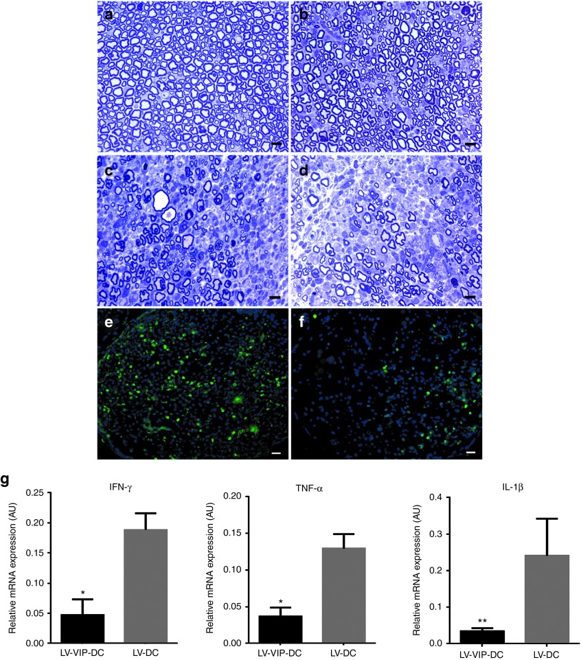
LV-VIP-DC treatment protects myelinated fibers and reduces inflammation in sciatic nerve. One-micrometer-thick cross-sections from midsciatic nerve segments before the disease onset at 16 weeks is shown in (a). Representative pictures of myelinated fibers from (b) LV-VIP-DC, (c) LV-DC, and (d) saline-injected groups at 25 weeks of age are shown. Scale bar = 10 µm. CD3+ T cells infiltration in midsciatic nerve samples of (e) LV-DC and (f) LV-VIP-DC groups at 25 weeks of age. Scale bar = 10 µm. For quantitative data see Table 2. Expression levels of proinflammatory cytokines in sciatic nerves from LV-VIP-DC injected and LV-DC groups from cohort 1 at 25 weeks of age (g). Total RNA was extracted from snap frozen sciatic nerve samples and analyzed for expression of proinflammatory cytokines TNF-α, IL-1β, and IFN-γ by using real-time quantitative PCR. These data were normalized to glyceraldehyde 3-phosphate dehydrogenase. Error bars represent standard error of the mean (n = 6–9 in each group; two-tailed t-test; *P < 0.05, **P < 0.01). AU, arbitrary units; DC, dendentric cell; IFN, interferon; IL, interleukin; LV, lentiviral vector; TNF, tumor necrosis factors; VIP, vasoactive intestinal polypeptide.

