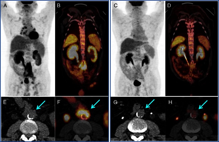
Fig. 3.
18F-FDG PET/CT guided the timely ureter recanalization in an IgG4-RD patient with retroperitoneal fibrosis and aorta involvement. The enlarged right renal pelvis with radioactive urine retention indicated severe ureteral obstruction, whereas the left side without radioactivity indicated complete obstruction (a and b). After D-J tube cannulation, the renal function was recovered bilaterally (c and d); the intense-uptake lesions were smaller and the intensity was significantly lower in response to the steroid treatment. The arrows show the aorta involvement beside the retroperitoneal fibrosis (e, f), which has a complete response to steroid-based treatment (g, h)

