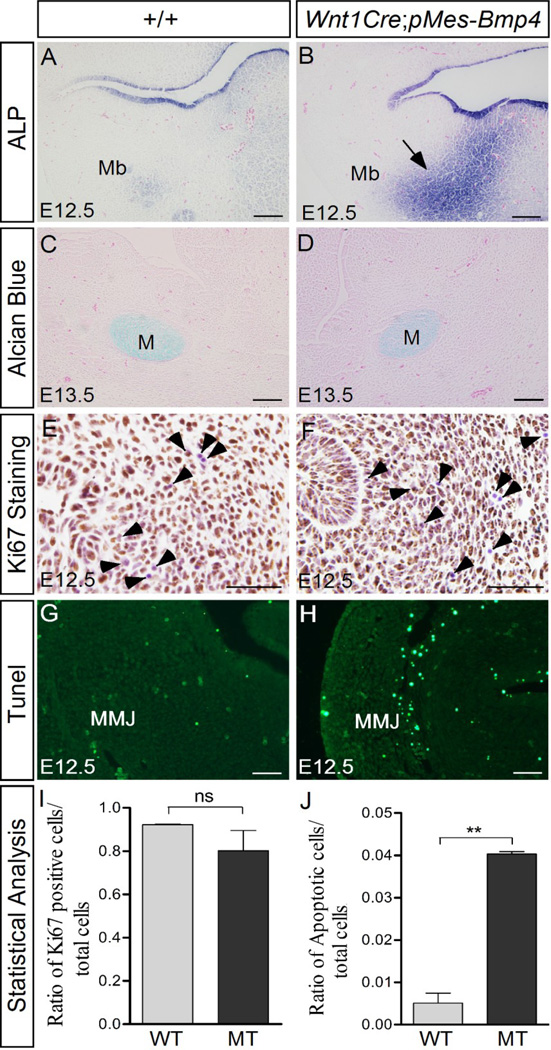
Figure 3. Wnt1Cre-driven Bmp4 expression causes abnormal osteoblast differentiation and apoptosis in the mandible.
(A, B) Alkaline phosphatase (ALP) staining reveals moderate osteoblast differentiation in the control mandible at E12.5 (A), but strong ALP activity in the mutant mandible mesenchyme (arrow in B). (C, D) Alcian blue staining results show comparable chondrocyte formation in the control and mutant mandible. (E, F, I) Immunostaining with anti-Ki67 antibody reveals a slight decrease of cell proliferation level in the mutant mandible. Arrowheads point to Ki67-negative cells that were counterstained by Hematoxylene (purple). (G, H, and I) Tunel assay shows abundant ectopic cell apoptosis in the mutant maxillo-mandibular junction. M, Meckel’s cartilage; Mb, mandible; MMJ, maxillo-mandibular junction. Scale bar = 100-µm in (A–D), and = 50-µm in (E–H).

