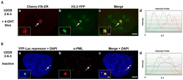
Figure 3.
Images of Regulatory Factor Organization at the Transgene Array.
(A) Histone H3.3-YFP (a) is strongly recruited to the activated transgene array in the ATRX-null U2OS cell line (2-6-3) in which replication independent histone H3.3 chromatin assembly is blocked. H3.3-YFP does not co-localize with the chromatin of the activated array marked by Cherry-tTA-ER (b). Arrows mark the location of the transgene array. Yellow lines in enlarged merge insets (c) show the path through which the signal intensities were measured in the intensity profiles (d). Asterisks mark the start of the measured line. Scale bar =5 0m. Scale bar in enlarged inset =1 0m.
(B) PML protein (b) is enriched at the inactive CMV-promoter regulated transgene array in the U2OS cell line (2-6-3), marked by YFP-lac repressor (a). DAPI DNA staining is shown in the merged images (a and c). (b)PML immunofluoresence staining was done with antibody PG-M3 (Santa Cruz Biotechnology).

