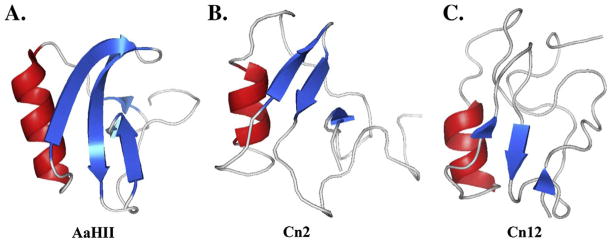
Figure 1. Three-dimensional structures of Na-ScTxs.

The structures shown are: α-NaScTx AaHII from Androctonus australis Hector (pdb 1SEG), β-NaScTxs Cn2 (pdb 1CN2) and Cn12 (pdb 1PEA) from Centruroides noxius Hoffmann). The structures were obtained from Protein databank (www.pdb.org) and were displayed with PyMOL (www.pymol.org). AaHII is the prototype of α-NaScTxs and Cn2 is a typical β-NaScTx; however, Cn12 is structurally a β-NaScTx, but it has a α-NaScTx effect. All these toxins have a three-dimensional structure highly conserved comprising an α-helix (red) and three or four-stranded anti-parallel β-sheets (blue).
