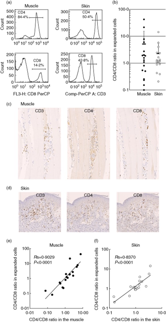
Fig. 1.

CD4/CD8 ratios in muscle- and skin-derived T cells in dermatomyositis (DM) patients. (a) Flow cytometric analysis (FCM) of expanded T cells. The numbers represent CD4+ or CD8+ cells. (b) The CD4 : CD8 ratio of expanded T cells from muscle lesions (closed circles, n = 18) and skin lesions (open circles, n = 18) of DM patients. Horizontal bars indicate mean ± standard deviation (s.d.). (c,d) Immunohistochemical staining of muscle and skin lesions with monoclonal antibodies against CD3, CD4 and CD8. Original magnification ×200. (e) Relationships between the CD4 : CD8 ratio of the original tissue-infiltrating T cells and that of expanded T cells from muscle lesions (closed circles) and skin lesions (open circles).
