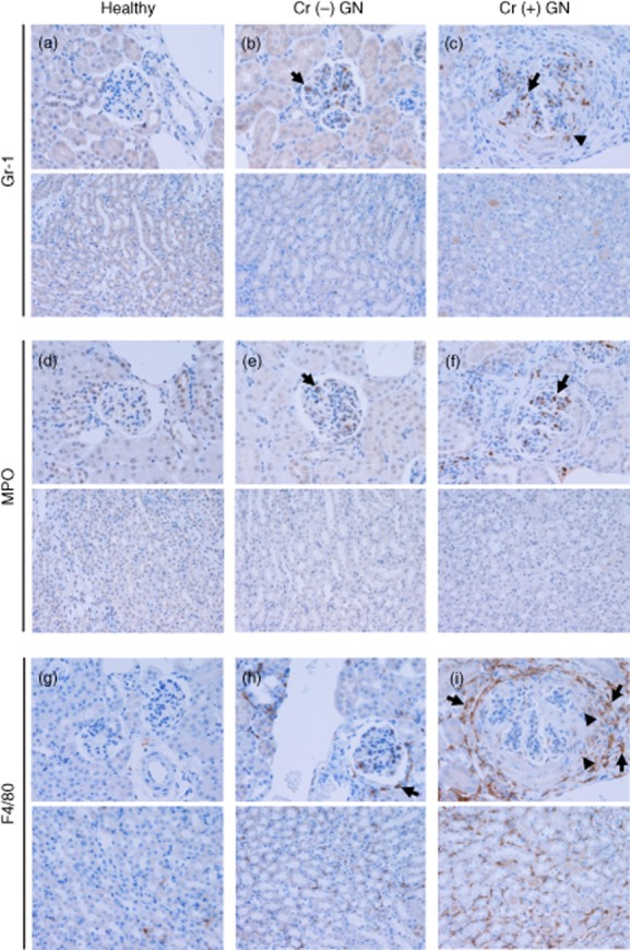
Fig. 2.

Distribution of Gr-1+ cells, myeloperoxidase (MPO+) cells and F4/80+ cells in healthy and glomerulonephritic glomeruli with crescents [Cr (+)] and without crescents [Cr (−)]. Upper photographs: cortical glomeruli, lower photographs: interstitium. (a–c) Gr-1+ cells were detected mainly within the glomerular tuft (arrows). Gr-1+ cells were also detected in crescents to a lesser extent (arrowhead), whereas they were rarely observed in the peri-glomerular area and in the interstitium. (d–f) As with Gr-1+ cells, MPO+ cells were found mainly in the glomerular tuft (arrows). They were detected occasionally in crescents, while they were rarely found in the peri-glomerular area and in the interstitium. (g–i) F4/80+ cells were present in the medullary and cortical interstitium in healthy mice. In mice with non-crescentic [Cr (−)] GN, an increased presence of F4/80+ cells was demonstrated in the peri-glomerular area (arrow) and in the interstitium. This was the case in mice with crescentic [Cr (+)] GN to a greater extent, and F4/80+ cells were also detected in crescents. F4/80+ cells in the interstitium and peri-glomerular area were F4/80high (arrows), while those in crescents were F4/80low (arrowheads). Original magnifications × 200. Numbers of mice: healthy, one; Cr (−) GN, three; Cr (+) GN, eight.
