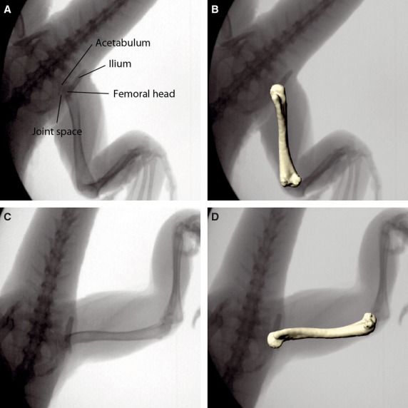
Fig. 8.

Femoral translation during in vivo locomotion. X-ray images of the hip joint configuration during the step cycle in touch down (A) and lift off (C) position; the same X-ray images with matched virtual femur model (B,D). Note that joint space is small and invariable. Minor femoral translation is apparent along the long-axis of the oval femoral head. This translation is considered in the scientific rotoscoping approach.
