
Fig. 6.
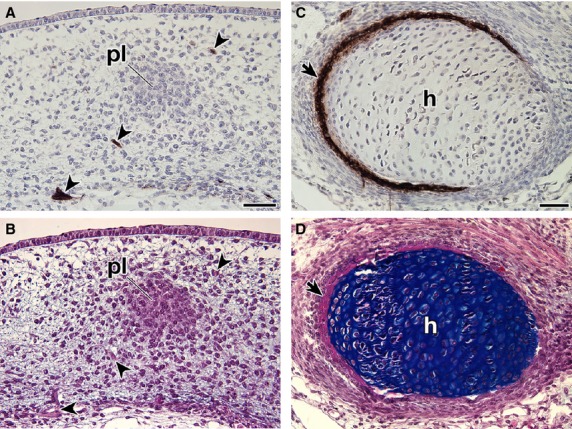
Expression of the HNK-1 epitope in Trachemys scripta embryos. Transverse sections of T. scripta embryos were either stained with HNK-1 and counterstained with hematoxylin (A,C) or simply stained with HE and then Alcian blue (B,D). (B,D) are adjacent sections to (A,C), respectively. (A, B) Higher magnification of the box (asterisk) in Fig. 5B, showing distribution of the HNK-1 epitope in the initial anlage of the plastral bones in the G stage 17 embryo. Note that the anlage is HNK-1-negative, whereas the peripheral nerves are strongly stained with the antibody (arrowheads). (C,D) Distribution of the HNK-1 epitope in the humerus (h) of the G stage 18 embryo. Note that the expression domain of HNK-1 overlaps the area of the intramembranous bone (arrows). Scale bars: 50 μm.