
Figure 2.
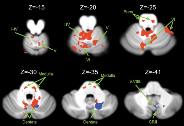
Connectivity maps in the cerebellum. Functional connectivity maps for the dorsal (red) and ventral (blue) dentate in the cerebellum (axial views; both presented at P < 0.00001). The dorsal network includes lobules I–IV, V, and VI in the anterior regions of the cerebellum. The ventral network is predominately made up of more posterior regions including crus II, but also extends into lobule VI. Images are oriented such that the right hemisphere is presented on the right. Roman numerals indicate the cerebellar lobules. CRII: crus II.