
Figure 3.
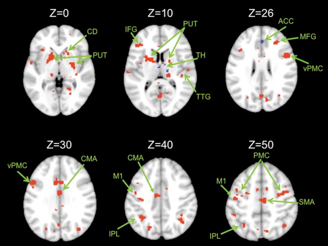
Connectivity maps in the whole brain. Functional connectivity maps for the dorsal (red) and ventral (blue) dentate seeds in the whole brain (axial views; both presented at P < 0.00001). The dorsal network consists primarily of motor and parietal regions, though there are also prefrontal regions included. The ventral network consists of the anterior cingulate cortex and the caudate. Images are oriented such that the right hemisphere is presented on the right. ACC, anterior cingulate cortex; CMA, cingulate motor area; CD, caudate; IPL, inferior parietal lobule; M1, primary motor cortex; MFG, middle frontal gyrus; PUT, putamen; SFG, superior frontal gyrus; TH, thalamus; TTG, transverse temporal gyrus; PMC, premotor cortex; d, dorsal; v, ventral. Unlabeled areas are extensions from the clusters presented in Table 2.