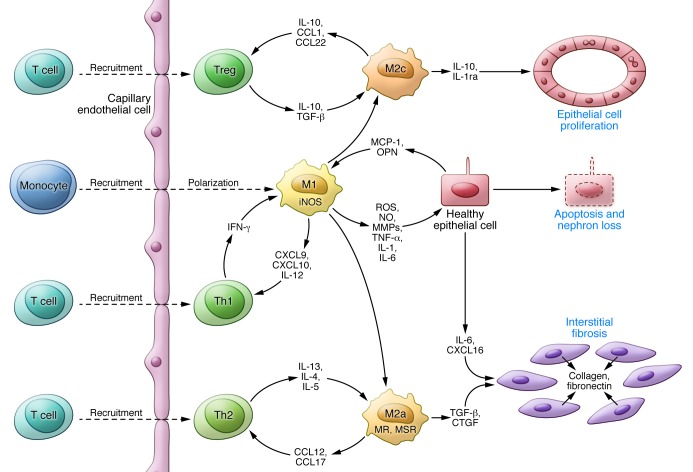
Figure 3. A model for the contribution of macrophages to PKD progression.
Activation of signaling pathways and transcription factors (e.g., STAT3, NF-κB) in cyst-lining cells stimulates the production and release of chemokines (e.g., MCP-1, osteopontin) attracting monocytes, promoting the polarization of invading monocytes and resident macrophages to a proinflammatory phenotype, and activating Th1 lymphocytes with further release of mediators and tissue damage. Opsonization of apoptotic cells by pentraxin-2 and secretion of IL-10 and TGF-β by immunosuppressive regulatory T cells promote the polarization of macrophages to a proproliferative phenotype, releasing antiinflammatory cytokines that induce cell proliferation. Incomplete epithelial healing, ongoing injury, and release of IL-4 and IL-13 by Th2 lymphocytes promote the polarization of macrophages to a profibrotic phenotype, releasing TGF-β and connective tissue growth factor (CTGF), which induces the differentiation of fibroblasts into collagen-secreting myofibroblasts.

