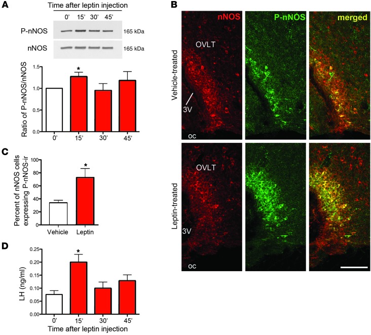
Figure 1. Leptin activates nNOS in the preoptic region and increases circulating LH levels.
(A) Representative Western blots for phosphorylated and total nNOS at the times indicated (in minutes) following leptin treatment (see Supplemental Figure 4 for full-length photographs of the Western blots). Leptin promotes the phosphorylation of nNOS acutely at 15 minutes. (B) Coronal sections of the OVLT, showing an increase in the percentage of nNOS cells expressing P-nNOS immunoreactivity (ir) 15 minutes after leptin stimulation. 3V, third ventricle; oc, optic chiasm. Scale bar: 100 μm. (C) Quantification of immunolabeling shown in B. (D) Circulating LH levels surge 15 minutes after leptin administration. *P < 0.05.

