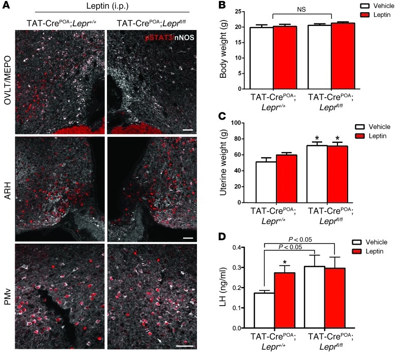
Figure 6. Site-specific deletion of LepR disrupts basal LH levels in female mice.
(A) Bilateral injections of TAT-Cre protein into the OVLT/MEPO prevented P-STAT3 45 minutes following peripheral leptin injection in the preoptic region (POA) of Leprfl/fl, but not in Lepr+/+, littermates. However, leptin was still able to induce P-STAT3 in caudal areas of the hypothalamus, such as the ARH and PMv, in TAT-CrePOA Leprfl/fl mice. Scale bar: 200 μm. (B) Body weight did not differ between TAT-CrePOA Lepr+/+ and TAT-CrePOA Leprfl/fl mice. (C) Lack of leptin signaling in the OVLT/MEPO resulted in higher uterine weight in TAT-CrePOA Leprfl/fl mice when compared with that of TAT-Cre–injected wild-type littermates. (D) Strikingly, basal levels of LH were increased in females lacking LepR signaling in the preoptic region when compared with those of wild-type mice injected with TAT-Cre. When injected with leptin, TAT-CrePOA Leprfl/fl mice were unable to respond further with a rise in LH levels. Scale bar: 50 μm. *P < 0.05.

