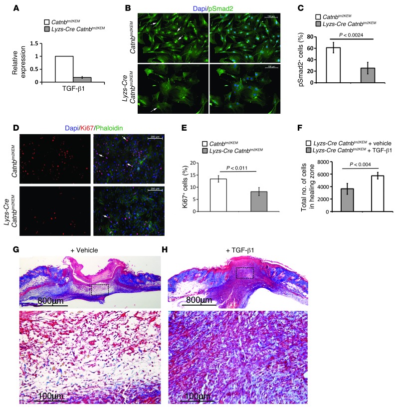
Figure 7. Macrophages lacking β-catenin induce less TGF-β1 signaling, and TGF-β1 partially rescues the wound phenotype of mice lacking β-catenin in macrophages.
(A) Quantitative RT-PCR analysis showing decreased TGF-β1 expression in β-catenin–deficient macrophages from Lysz-Cre Catnbtm2KEM ROSA-EYFP mice compared with that seen in their control littermates. (B) pSmad2 staining of fibroblasts that were exposed to either control (top panels) or β-catenin–deficient macrophages (bottom panels), indicating less pSmad2-positive cells in fibroblasts that were exposed to macrophages lacking β-catenin (quantified in C). Arrow shows pSmad2-positive cells. Scale bars: 100 μm. (D) Ki67 staining of fibroblasts that were exposed to either control macrophages (top panels) or to β-catenin–deficient macrophages (lower panels), indicating less Ki67-positive cells in fibroblasts that were exposed to macrophages lacking β-catenin (quantified in E). Arrow shows Ki67-positive cells. Scale bars: 200 μm. (F) Cell density quantification shows a significant increase in the number of cells in healing wounds in mice treated with TGF-β1 compared with those treated with vehicle. (G and H) Representative histology of a wound from a mouse treated with vehicle or TGF-β1 whose macrophages lacked β-catenin. Image in G is magnified in the lower panel; scale bars: 800 μm; 100 μm. Image in H is magnified in the lower panel; scale bars: 800 μm; 100 μm. Data represent the mean ± 95% CI of 7 mice.

