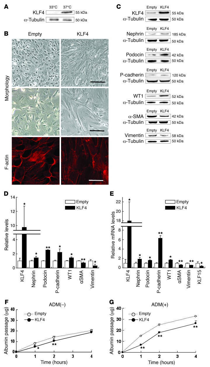
Figure 5. KLF4 expression causes morphological changes and induces epithelial cell markers in cultured podocytes.
(A) Western blot analysis of KLF4 expression in undifferentiated podocytes cultured at 33°C or differentiated podocytes cultured at 37°C. (B) Representative photomicrographs of (upper/middle panel) cell morphology at high/low confluence and (lower panel) immunofluorescence labeling with F-actin in KLF4-overexpressing podocytes and empty vector–transfected controls. Scale bars: 100 μm (upper panel); 50 μm (middle panel); 10 μm (lower panel). (C) Representative Western blots and (D) quantification of epithelial or mesenchymal markers expression in KLF4-overexpressing podocytes and controls (n = 4). (E) Real-time RT-PCR analysis of mRNA expression in KLF4-overexpressing podocytes and controls (n = 6). (F and G) In vitro permeability of FITC-labeled albumin through podocyte monolayers in KLF4-overexpressing podocytes and controls without (F) or with (G) ADM (0.3 μg/ml) for 48 hours (n = 6). The passage of albumin was measured at indicated hours after FITC-albumin administration. *P < 0.05; **P < 0.01 vs. controls.

