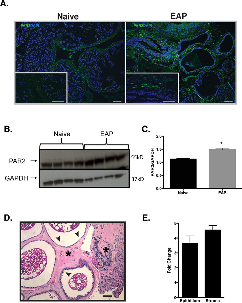
Fig. 2. PAR2 is elevated in the prostate of EAP mice.

(A) Representative immunofluorescence images of the prostate showed that NOD mice with EAP have elevated PAR2 expression (green) in the stroma (insets) compared to naive cohorts. DAPI is labeled blue and bars represent 50 microns. (B) Immunoblot and (C) densitometry data confirmed that the prostate from EAP mice (n=4) up-regulate PAR2 expression, compared to naive (n=4). (D) Representative H&E stained section of a prostate from a NOD mouse with EAP were used to microdissect epithelium (arrows) and stroma (asterisks) separately. (E) Real-time PCR analysis showed increased in mRNA for PAR2 in the stroma and epithelial layers of mice with EAP (pooled). Data was normalized to L-19 and expressed as fold change respective to naive. (*) denotes p<0.05. Real-time PCR experiments were performed three times.
