
Figure 1.
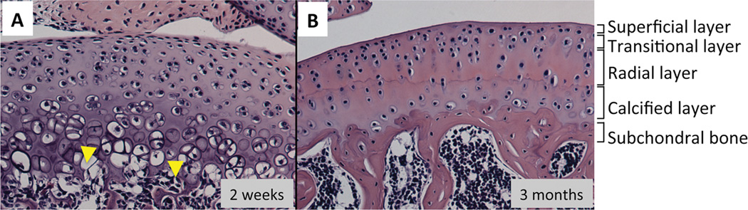
Comparison of histology between developing and mature articular cartilage. The longitudinal section of tibial plateau prepared from 3-weeks old (A) or 3-months old (B) C57BL6 mouse were stained with hematoxylin and eosin. A, The developing articular cartilage show vascular invasion at the bottom (arrow). B, The mature articular cartilage has superficial, tangential, radial and calcified layers, and subchondral bone lies below the calcified zone.